
Introduction
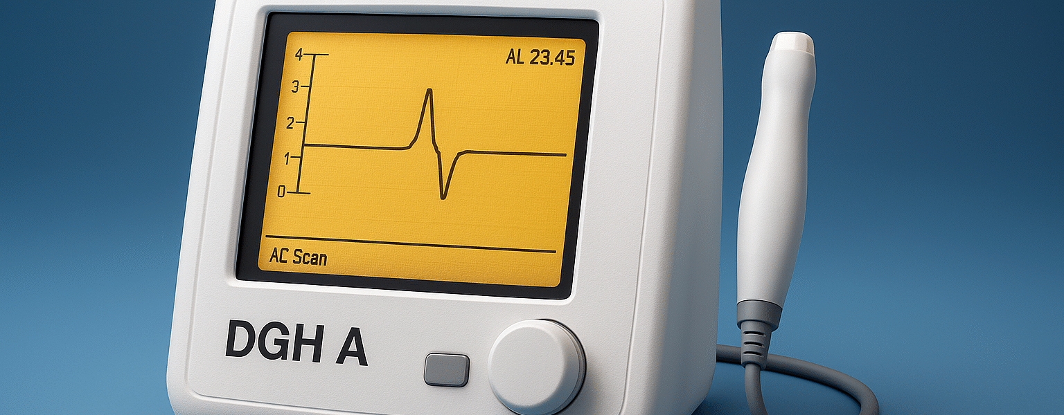
The DGH A and known under the Scanmate A (DGH 6000) is a medical device employed by eye physicians. This is an A-Scan ultrasound machine. It implies that it enters sound pulses into the eye. Then it writes the way back echoing of the sound. Based on echoes, the machine detects components of the eye including the eye length and lens thickness.
These figures are extremely significant. Optical practitioners adopt them to select the appropriate artificial lens in cataract surgery. They are also useful in the checking of certain eye issues. In this paper you will find out:
- The DGH A (Scanmate A) what it is and how it works.
- Key features and specs.
- How doctors use it in clinics.
- Safety tips and rules.
- Its comparison with other instruments and novel trends of eye biometry.
I shall not use complicated languages or long sentences, hence this will be read quickly and easily.
What is an A-Scan ultrasound?
The simplest type of ultrasound applied to the eye is an A-Scan. It reflects itself in the form of spikes on a screen. The eye is punctured with spikes on its surfaces. As an example, the cornea, lens and retina. The spike distance informs the doctor of the length of the eye. This is called axial length.
A-Scan assists in determining the strength of an implanted lens (an IOL) of the cataract patients. It is also applied in instances where optical equipment is too cloudy to use on the eye. A-Scan may be carried out in two forms which are: contact (applanation) or immersion. Eye clinics are taught and utilize both methods.
Who makes the DGH A (Scanmate A)?
DGH Technology, Inc. produces the DGH A. The scanner is marketed as the Scanmate A or DGH 6000 Scanmate-A. DGH is a manufacturer of a couple of ophthalmic ultrasound devices, such as A-Scan and B-Scan.
What the DGH A (Scanmate A) does — simple list
- Assesses the axial eye length (length).
- Measures anterior chamber depth (space between cornea and lens).
- Measures lens thickness.
- Helps is used to compute IOL power in cataract surgery.
- Stores loads the measurements of exports to a computer.
Key features of the DGH A (Scanmate A)
Scanmate A is also referred to as being portable and accurate. The key characteristics of it in simple words are as follows:
- Portable design: It attached to a laptop or a computer and can be carried around between exam rooms. This is useful with the small clinics or mobile teams.
- Clear measurement software: The equipment employs computerized software to process real time waveforms. The software does provide feedback in order to assist the user make good measurements. DGH Technology
- High resolution: It displays fine detail of measurements (resolution to 0.01 mm in other models). This assists in coming up with more precise IOL calculations.
- Multiple modes: The Scanmate is capable of contact (applanation) and immersion A-Scan. In some cases, the way to be more accurate is through immersion.
- IOL calculations: The unit has up to date IOL formulas. That assists the surgeon in the selection of the right lens power.
How the DGH A (Scanmate A) works — the basics
- Probe sends sound pulses.
The physician carries a thin probe towards the eye. It transmits low powered ultrasound pulses. - Echoes return.
The pulses bounce back from eye structures. The device records the echoes. - Waveform appears.
The echoes are reflected as spikes in the screen. The spikes represent each surface within the eye. - Software measures distances.
The interval between spikes is changed to distances (such as axial length). - IOL power is calculated.
The software assists in calculating the power of the lens to be used in surgery using these distances and a selected formula.
Measurement modes: Contact vs Immersion
- Contact (Applanation):
The investigator rubs the probe against the eye. This method is quick. However, touching the eye a bit can be used to reduce the reading. That may have an impact on IOL power calculations. - Immersion:
A little shell or bath is applied between the probe and the eye with saline. There is no physical contact of probe with the cornea. It will minimize the compression of the cornea and may be more precise in the eyes of some individuals. Numerous centers choose to immerse difficult cases.
The Scanmate A is compatible with the two techniques. The user selects the most appropriate approach to every patient.
Why axial length (AXL) is so important
An IOL is more sensitive to the single most significant number which is the axial length of the eye. Even the slightest mistake during the axial length may alter the outcome of the surgery vision. Incidentally, a 0.1 mm difference in the axial length can cause a refractive outcome to move an average of 0.25 to 0.3 diopters- enough to cause the patient to require glasses. That is why it is important to have valid A-scan measurements.
Typical DGH A (Scanmate A) specs (what clinics care about)
The following are standard values that are given in product descriptions and documentation. The real numbers differ depending on the model and program (.)
- Axial length range: ~15.0 mm to 40.0 mm.
- Anterior chamber depth (ACD): 2.0 mm -6.0 mm.
- Lens thickness (LT): ~2.0 mm to 7.5 mm.
- Resolution: 0.01 mm reported on some listings.
- Transducer frequency: commonly A-Scan transducer probes have about 10 MHz.
- Types of measurement: Immersion and contact.
The device manual by DGH Technology contains detailed numbers. Safety and set up are also described in the manual.
Who uses the DGH A (Scanmate A)?
- Ophthalmologists (surgeons of an eye).
- The clinics with biometry are those operated by optometrists.
- Surgery centers where cataract is intended to be carried out.
- Mobile eye units which require mobile devices.
The device is useful in clinics in which the optical biometry (such as optical interferometry devices) is not applicable due to the cataract density or uncooperative eye.
Training and user skills
An adequate A-Scan reading requires practice. The device assists in providing direction. Yet the user must:
- Hold the probe steady.
- Make certain that there is alignment along the visual axis of the eye.
- Learn to use immersion or contact.
- Check waveforms- check similar spikes and repeat measures.
DGH offers software instructions and manuals. The use of short hands-on training by the staff in many clinics is also common.
Safety notes and regulation
Ophthalmic ultrasound should be applied with caution. The eye is sensitive to ultrasound energy. Eyewear devices must be safe. In United States, FDA regulates ultrasound equipment and offers advice on safe application of ophthalmic ultrasound. To have safe exams the training and going through the device manual is a prerequisite. United States Food and Drug Administration+1.
Comparing DGH A to other A-Scan devices
It has a number of A-Scan brands. Some are small desktop units. Others connect to a PC. What is the comparison of the DGH Scanmate A?
- Portability: Scanmate A is highly portable and can be connected to a computer. This is beneficial to small clinics or mobile services.
- Software characteristics: DGH focuses on the analysis of the real-time waveform and the instruction of the user. This assists in making good measurements within a shorter time.
- Accuracy: Repeatability values given (e.g. +-0.03 mm SD in immersion of some) are reputed to be very accurate when used properly. Certain values are model and technique specific.
Many of the eyes can be more accurate with optical biometers (such as the IOL Master or Lenstar). However, A-Scan such as DGH device is a sure option when the optical paths are not available (dense cataract).
Common problems and troubleshooting
- Bad waveform quality: Can be as a result of bad probe alignment, or excessive probe pressure. Attempt to re-align or change to immersion.
- Problematic inconsistencies: Get multiple measurements and mean the best results. Make use of guidance facilities of the device.
- Electrical or software problems: Use the manual and use the support of DGH when necessary. EMC warnings and suggested computer configurations are pointed out in the operator manual.
Practical tips for clinics
- The A-Scan should be tested and calibrated regularly.
- Immersion should be used in difficult or thick cataracts in order to enhance accuracy.
- Train operators are trained on the software prompts and checking of the waveforms of the device.
- Use optical biometry in case of A-Scan results.
- Record the measurements and the IOL formula adopted on each patient. Enhanced Medical Services+1
Future of A-Scan and DGH devices
The A-Scan technology is still relevant. A-Scan remains essential though optical biometers are improving at the point that optics are not useful. DGH and other makers upgrade the software and add features. The portable devices which connect to computers will continue to serve the small clinics and outreach programs.
Among the trends, there is improved waveform analysis, cloud data storing, and easier interconnection with surgical planning software. The changes increase the speed of the work and assist surgeons in achieving improved results.
Conclusion
DGH A (Scanmate A) is a reliable A-Scan ultrasound eye care. It determines the eye length, thickness of the lenses and the chamber depth. These numbers are used by doctors to plan cataract surgery and also in assessing eye health by using optical tools when optical tools fail.
The Scanmate A is portable. It provides real time analysis and accommodates contact and immersion methods. As it is well trained and the technique applied, it produces precise and repeatable measures. It can enhance patient outcomes and surgical planning in the clinics that utilize the tool.


Couldn’t have said it better myself.